
Technology
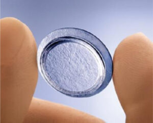
Prokera®
PROKERA® is a therapeutic device used by eye doctors around the world to protect, repair and heal damaged eye surfaces. PROKERA® is made by clipping a piece of amniotic membrane tissue in between two rings made out of a clear, flexible material.
What is amniotic membrane tissue?
Amniotic membrane is part of the placenta and is the tissue closest to the baby throughout development in the womb. Amniotic membrane protects the baby from any harm and has natural therapeutic actions which help the baby develop. The tissue has healing properties that aid in ocular surface repair.
What does PROKERA® do?
The amniotic membrane tissue in PROKERA® has natural therapeutic actions that help damaged eye surfaces heal. Eyes treated with PROKERA® have quicker healing, less pain, less scarring, and less inflammation. The amniotic membrane in PROKERA® is thin and clear like the tissue on the surface of your eye and protects your eye’s damaged tissue while inserted.
What does PROKERA® treat?
PROKERA® is used by eye doctors to treat eye diseases such as keratitis, corneal scars, chemical burns, corneal defects, partial limbal stem cell deficiency and many other ocular surface diseases with inflammation.
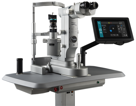
Yag Laser
Yag laser technology for two types of office laser surgeries.
- Yag Laser Iridotomy
- Yag Laser Capsulotomy
Yag Laser Iridotomy
Narrow angles may be a precursor to angle-closure glaucoma, the kind of glaucoma that can have a sudden, painful onset or a slow unrelenting downhill course.
The best time to prevent the damage that angle-closure glaucoma can cause is to treat it with a laser iridotomy before the actual disease sets in. This is preventative medicine at its best.
Although not everyone with narrow angles actually develops glaucoma, careful evaluation of the angle structure can identify who is at greatest risk. The angle structure is determined by an examination called gonioscopy which is performed with a special contact lens called a gonioprism.
By performing a laser iridotomy, your doctor is trying to save you from the risk of acute angle-closure glaucoma.
Yag Laser Capsulotomy
The natural lens has a cellophane-like outer lining called the capsule. During cataract surgery the back membrane of the natural lens (posterior capsule) is left in place to support the artificial lens implant. The posterior capsule is normally clear, however, 3 out of 4 people who have cataract surgery will eventually develop a cloudiness of this membrane.
The cloudiness which can develop months or years later is a result of scarring (a normal healing response) and can interfere with vision in ways similar to the original cataract. If the clouding of the posterior capsule interferes with your vision, your doctor may suggest opening the capsule to restore the best sight that can be achieved.
This is done with a procedure called YAG laser capsulotomy, whereby your doctor uses a laser beam to make a tiny hole in the posterior membrane to let light pass through and restore clear vision. Although, the laser procedure requires close and precise focusing by the ophthalmologist, for the patient the technique is a painless, outpatient procedure and is never part of the original cataract operation.
There is a common misconception that cataracts are removed by a laser. This stems from the fact that 3 out of 4 people who have cataract surgery eventually need YAG laser surgery to remove the cloudy membrane which is often referred to as a secondary cataract.
A YAG laser capsulotomy is a surgical procedure; however, the risks of a serious complication resulting from this procedure are about 1/100th of the risks associated with a regular cataract operation. The most serious risk is of retinal detachment which at it’s onset displays itself as a black curtain coming over the eye affecting the side vision from any direction. The occurrence of retinal detachment may also be associated with the appearance of flashing lights. If you experience either of these symptoms you should contact your eye doctor immediately.
SLT – The Gentle Alternative for Glaucoma
A New Hope For Glaucoma
Gentle and painless, SLT is a simple laser procedure that takes just five minutes to perform at Eye Care Northeast, PC. Not only can SLT effectively reduce elevated IOP but it can also free you from the financial and lifestyle burdens of glaucoma medications. Also known as Selective Laser Trabeculoplasty, SLT is a simple, yet highly effective laser procedure that reduces intraocular pressure. It is performed at Eye Care Northeast, PC, and typically takes no more than five minutes.
How Does SLT Work?
SLT stimulates a natural healing response in the body. The laser therapy uses short pulses of low-energy light to target the melanin, or pigment, in specific cells of the eye. In response, the body’s natural healing mechanisms go to work to rebuild these cells. This rebuilding process improves drainage and lowers intraocular pressure.
Does SLT hurt?
No. SLT is painless, and there are no side effects to worry about.
Some Statistics About Glaucoma
- It is estimated that over 3 million Americans have glaucoma but only half of those know they have it.
- In the U.S., more than 120,000 are blind from glaucoma, accounting for 9% to 12% of all cases of blindness.
- Glaucoma is the second leading cause of blindness in the world, according to the World Health Organization.
- Other high-risk groups include: people over 60, family members of those already diagnosed, diabetics, and people who are severely nearsighted.
- Estimates put the total number of suspected cases of glaucoma at over 60 million worldwide.
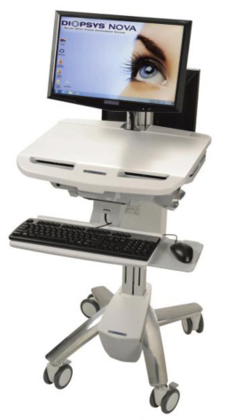
Early detection means better patient outcomes.
Diopsys is the leader in providing ophthalmologists with objective, functional information about the vision system to aid in the early detection of vision disorders and patient management through ERG and VEP vision testing.
The CIRRUS HD-OCT 6000
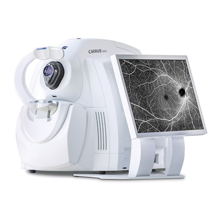
The CIRRUS HD-OCT 600 is an advanced eye imaging device that helps doctors diagnose and manage various eye conditions. It provides detailed images of the retina, which is essential for monitoring glaucoma and retinal diseases, assessing the retina before cataract surgery, and examining the cornea for diseases.
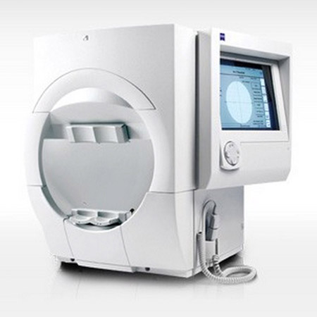
The Zeiss Clarus 500
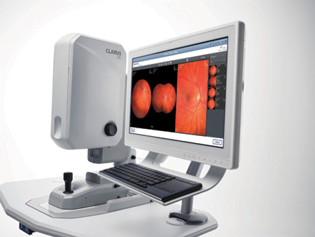
The Zeiss Clarus 500 is a high-tech camera used by eye doctors to capture detailed, high-resolution images of the retina. This helps in accurately diagnosing and monitoring eye conditions such as retinal diseases, glaucoma, and other issues. The clear, wide-field images make it easier for doctors to provide comprehensive eye care and ensure your vision health is well-managed.
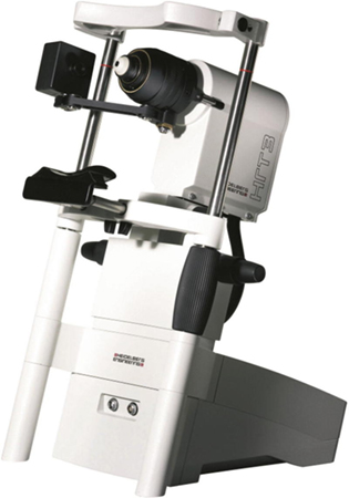
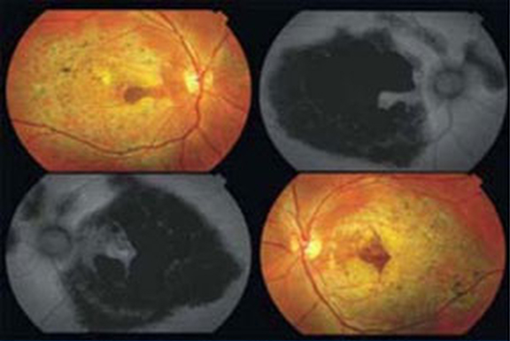
Heidelberg Retinal Tomography
The Heidelberg Retinal Tomograph (HRT) set the standard for analyzing 3D structural changes to the retina, especially in examining the optic nerve head. The secret of treating Glaucoma is catching it early when it is manageable.
These 3D images and the associated software lets us at Eye Care Northeast predict the development of Glaucoma and when to start treatment. The Heidelberg HRT also allows us to evaluate structural changes associated with other retinal conditions such as retinal edema, diabetic retinopathy, macular degeneration as well as many neurological problems such as brain tumors and Multiple sclerosis.
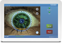
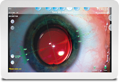
The Verion Image Guided System
As the leader in the global refractive cataract surgery category, Alcon continues to introduce the latest advancements in surgical innovations and technologies designed to optimize and improve the refractive cataract procedure.
The Verion Image Guided System is designed to improve accuracy and minimize error through:
- Imaging: The Verion Reference Unit performs almost all critical measurements in a single step, creating a high-resolution, digital reference image of the eye.
- Planning: The Verion Reference Unit combines multiple, proven algorithms to help surgeons determine the optimal IOL – including advanced technology IOLs – and lens power for patients with and without astigmatism.
- Guidance: The Verion Digital Marker provides automated centration and incision positioning in both LenSx Laser and traditional procedures. The system eliminates the need for manual toric eye markings prior to surgery, and also provides centration and alignment guidance for multifocal and toric IOLs, respectively.
Once the Verion Image Guided System has captured the reference image and helped generate a surgical plan, it integrates the surgical plan in the OR via a tracking overlay, allowing surgeons to see all incisions and alignment in real time.

Zeiss Veracity and Callisto patient IOL calculation and verification systems
VORACITY and CALLISTO by Zeiss which results in more accurate IOL power calculation, toric alignment and allows for greater customization of the surgery to each patient’s individual needs, providing them with the best possible vision. The combination of these technologies, along with other pre-operative diagnostic equipment such as the IOL Master, allows for precise IOL or lens implant calculations, thus more refined IOL/lens implant selection. With today’s incredible diagnostic and lens implant technology, most patients undergoing cataract surgery have incredibly precise visual outcomes that were only a dream a decade ago.
Zeiss Humphrey Field Analyzer 3
The Heidelberg Edge Perimeter (HEP) performs innovative functional testing of the visual system.
Another year has passed and it is time for your visual field test, a.k.a. “the clicky test” that frazzles many glaucoma patients and can feel like a waste of time. The office staff reassures you that it is a necessary part of your exam and you cooperate to the best of your ability. But why do you need to take this test?
The visual field test is a subjective measure of central and peripheral vision, or “side vision,” and is used by your doctor to diagnose, determine the severity of, and monitor your glaucoma. The most common visual field test uses a light spot that is repeatedly presented in different areas of your peripheral vision.
Stage-Specific Perimetry
Visual field testing remains the main diagnostic tool to provide information about a patient’s visual function, quality of vision and quality of life. Functional testing supports clinicians in early disease detection, staging of disease or trend analysis in progressive glaucoma.